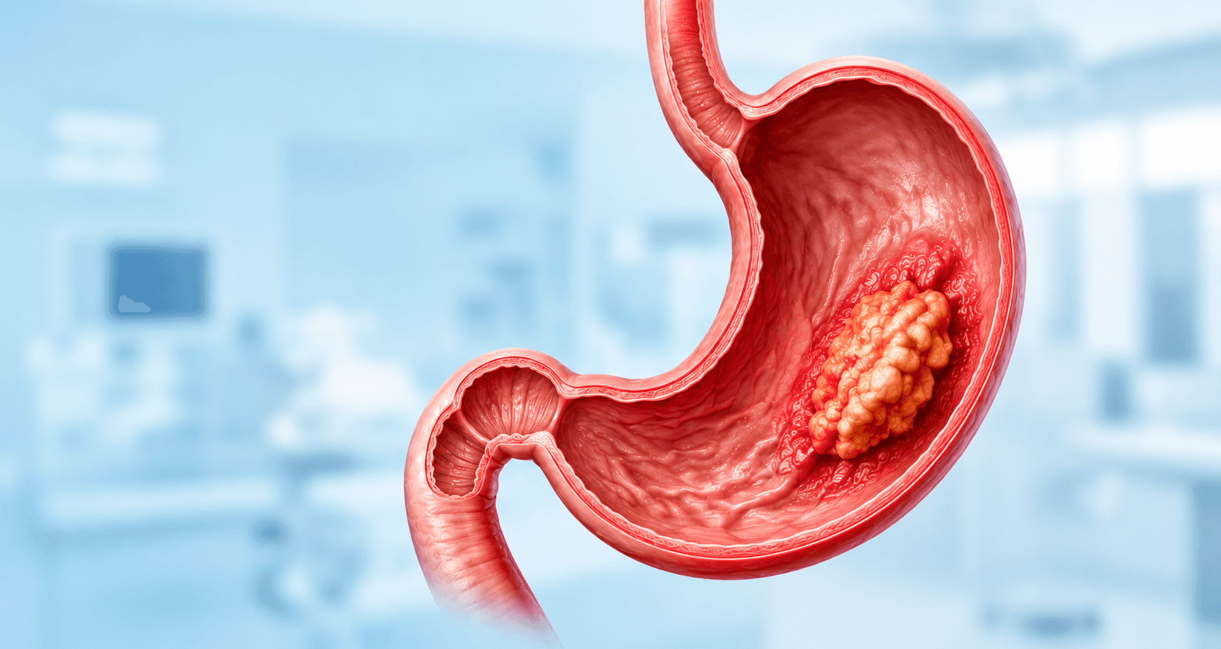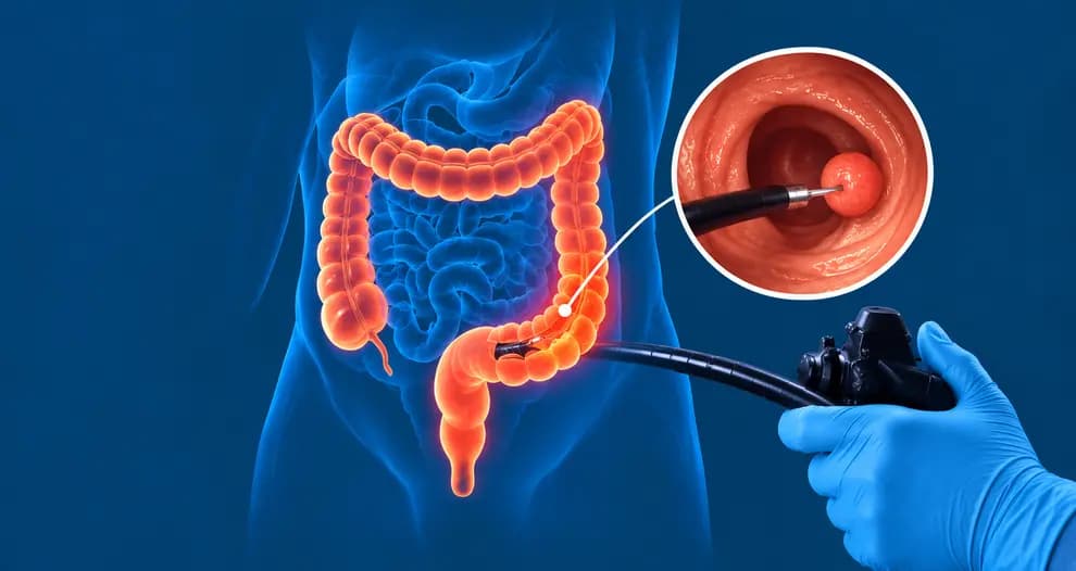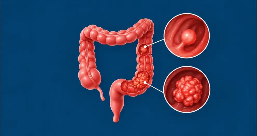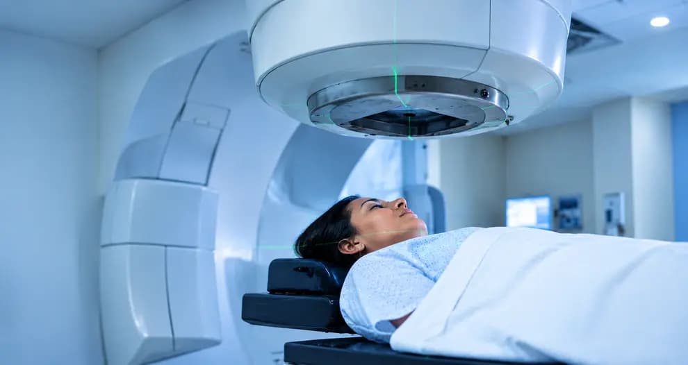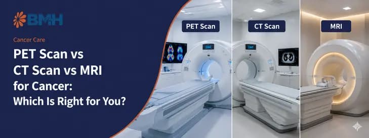

PET scans, CT scans, and MRI detect cancer in fundamentally different ways: CT scans map internal anatomy using X-rays, MRI provides highly detailed soft tissue imaging through magnetic fields, and PET scans reveal abnormal cellular activity using radioactive tracers.
A National Institutes of Health-backed meta-analysis of 38 studies found whole-body PET/MRI achieved 93% sensitivity and 92% specificity for malignancy detection, highlighting why no single scan fits every cancer case. The right imaging method at the right stage can significantly influence diagnosis, treatment planning, and patient outcomes.
What Are PET Scans, CT Scans, and MRI and How Do They Work?
Although PET scans, CT scans, and MRI are often grouped together under “cancer imaging,” they work in completely different ways and answer different clinical questions. Some scans show the physical structure of a tumour, while others reveal how actively cancer cells are behaving inside the body.
Understanding how each technology works makes it easier to see why doctors may recommend one scan over another or sometimes use multiple imaging methods together during diagnosis, staging, or treatment monitoring.
CT Scan (Computed Tomography)
A CT scan uses rotating X-ray beams and computer processing to create detailed cross-sectional “slice” images of the body. Unlike a standard X-ray, CT imaging captures multiple angles rapidly, allowing doctors to examine organs, bones, blood vessels, and tumours in far greater detail.
CT scans are widely used in cancer detection because they are fast, accessible, and highly effective for imaging the chest, abdomen, pelvis, and bone structures. Most scans take only 5–15 minutes.
In many cancer cases, an iodine-based contrast dye is injected into a vein before the scan. This contrast helps highlight blood vessels, tumour boundaries, inflammation, and abnormal tissue growth more clearly. Patients with reduced kidney function, an iodine allergy, or certain thyroid conditions may need a modified protocol or a different scan, so always share your medical history with the imaging team.
CT scans primarily produce anatomical or structural images. In simple terms, they show where a tumour is located, how large it is, and whether nearby organs appear affected. Outside oncology, CT is the workhorse of emergency imaging. Within minutes it can reveal fractures, internal bleeding, stroke, and trauma injuries, which is why it is usually the first scan ordered in an emergency.
MRI (Magnetic Resonance Imaging)
MRI uses powerful magnetic fields and radio waves to generate highly detailed images of soft tissues inside the body. Unlike CT scans, MRI does not use ionising radiation. MRI scans typically take longer, around 30–60 minutes (multi-sequence studies can run longer, up to about 90 minutes), and the machine can be loud and enclosed, which may feel uncomfortable for some patients.
In certain cases, especially for children or claustrophobic patients, mild sedation may be recommended.
MRI works by detecting how hydrogen protons in body water behave inside a magnetic field. Because different tissues hold different amounts of water, MRI separates them with exceptional contrast. A single session can also capture several image sequences that show tissue composition, cell density, blood supply, and diffusion, which helps both diagnosis and treatment planning.
MRI is considered the gold standard for evaluating soft tissue structures. It is especially valuable for detecting and assessing:
- Brain tumours
- Spinal cord abnormalities
- Prostate cancer
- Pelvic cancers
- Liver lesions
- Soft tissue invasion around tumours
While CT scans excel at speed and broader body imaging, MRI provides significantly better soft tissue contrast, helping doctors distinguish between healthy tissue, inflammation, and tumour involvement with greater precision. Like CT, MRI produces anatomical images, but with much finer soft tissue detail. Beyond cancer, MRI is also the preferred scan for nerves, ligaments, tendons, joint injuries, and conditions such as multiple sclerosis and stroke.
PET Scan / PET-CT (Positron Emission Tomography)
PET scans work very differently from CT or MRI because they focus on cellular activity rather than anatomy alone.
Before the scan, patients receive a small injection of a radioactive tracer — most commonly FDG (fluorodeoxyglucose), a glucose-like substance. Since cancer cells usually consume glucose much faster than normal cells, areas with high metabolic activity appear brighter or “light up” on the scan.
This allows PET imaging to identify potentially active cancer cells even before structural changes become clearly visible on CT or MRI.
Modern PET imaging is almost always combined with CT in a PET-CT scan. This combination allows doctors to see both:
- Functional activity (from PET)
- Precise anatomical location (from CT)
The actual scan usually takes 30–45 minutes, but patients first spend approximately 45–60 minutes in a tracer uptake waiting room after injection. In total, plan for about 2 hours at the centre; some centres quote up to 3 hours once registration and preparation are included. The tracer is short-lived. FDG is labelled with fluorine-18, which has a half-life of roughly 110 minutes, so its radioactivity fades quickly and drinking water after the scan helps flush it from the body. Although this article focuses on cancer, PET is also used to evaluate brain disorders such as Alzheimer’s disease and epilepsy, and to assess heart disease.
Choosing the right cancer scan becomes much easier once you compare what each imaging method is actually designed to detect.
PET Scan vs CT Scan vs MRI for Cancer: Quick Comparison
While all three imaging methods play an important role in cancer diagnosis and treatment planning, they are designed to answer very different medical questions. CT scans focus on structural abnormalities, MRI provides deeper soft tissue clarity, and PET-CT reveals how actively cancer cells are functioning inside the body.
The comparison below breaks down the key differences to help you quickly understand when each scan is typically preferred.
| Feature | CT Scan | MRI | PET-CT |
|---|---|---|---|
| Technology | X-ray (ionising radiation) | Magnetic field + radio waves (no radiation) | Radioactive tracer + CT (ionising radiation) |
| What it shows | Anatomy (structure) | Anatomy with superior soft tissue detail | Metabolic activity + anatomy |
| Best for | Chest, abdomen, bones, fractures, emergency staging | Brain, spine, prostate, liver, pelvic tumours | Whole-body cancer staging, recurrence detection, treatment response |
| Scan duration | 5–15 minutes | 30–60 minutes | 2–3 hours total (including tracer uptake time) |
| Radiation | Yes (low dose) | No | Yes (low dose) |
| Contraindications | Iodine allergy, kidney disease (with contrast) | Metal implants, pacemakers, claustrophobia | Uncontrolled diabetes, pregnancy |
| Cost (relative) | Lowest | Moderate–High | Highest |
| Availability | Widely available | Widely available | Usually limited to specialist imaging centres |
| Earliest change detected | Structural change (anatomy) | Structural change (soft tissue detail) | Metabolic change, often before anatomy changes |
| Contrast / agent used | Iodine-based contrast (optional) | Gadolinium-based contrast (optional) | Radioactive tracer (FDG) injected |
| Claustrophobia concern | Low (short, open ring) | Higher (longer, narrower tunnel; wide-bore scanners and sedation can help) | Low |
| Emergency use | Yes, first choice for trauma, bleeding, fractures | Limited | No |
| Suitable for repeat scans | Limited by cumulative radiation dose | Yes, no radiation | Limited by radiation dose |
Also Read: Targeted Therapy vs Chemotherapy: How to Choose the Right Treatment
The real difference between PET scans, CT scans, and MRI becomes clearer when you look at how doctors use them across different cancer types and treatment stages.
Which Scan Is Used for Which Cancer and When?
No single imaging test is considered “best” for every cancer. Doctors choose between CT, MRI, and PET-CT based on the tumour location, the information needed, how urgently results are required, and whether they need to evaluate structure, spread, or metabolic activity.
The breakdown below explains where each scan is most commonly used in real-world oncology practice.
Quick Guide: Which Scan for Which Cancer?
| Cancer / situation | Often used first | Often added | Why |
|---|---|---|---|
| Lung cancer | CT chest | PET-CT for staging | CT is better than MRI for lung tissue and small nodules; PET-CT shows whether nodes or distant sites are active. |
| Brain tumour | MRI | PET in selected cases | MRI shows how a tumour relates to critical brain areas; PET can help tell recurrence from radiation necrosis when MRI is inconclusive. |
| Spinal lesions | MRI | CT for bone detail | MRI shows the spinal cord and nerves; CT shows fine bone structure. |
| Prostate cancer | MRI (local staging) | Further imaging if spread is suspected, as your doctor advises | Diffusion-weighted MRI shows the extent of tumour within and around the gland. |
| Breast cancer | Mammography; MRI for high-risk patients such as BRCA1/2 carriers | PET-CT for advanced staging | MRI adds detail in dense or high-risk breasts and can assess response to pre-surgical chemotherapy. |
| Lymphoma | PET-CT | CT | PET-CT is standard for staging and end-of-treatment response in most lymphoma subtypes. |
| Head & neck, oesophageal | PET-CT for staging | CT or MRI for local extent | PET-CT detects nodal and distant spread; CT or MRI define local tumour boundaries. |
| Liver tumours | CT or MRI | MRI to characterise unclear lesions | MRI helps distinguish benign lesions from hepatocellular carcinoma when CT is non-specific. |
| Rectal, cervical, endometrial cancer | MRI (local staging) | CT or PET-CT for spread | MRI shows local invasion and surrounding structures before surgery or chemoradiation. |
| Cancer of unknown primary | PET-CT | MRI or CT as needed | PET-CT helps locate the source when metastases are found but the original tumour is not. |
This is a general guide. Your oncologist chooses the scan based on your symptoms, test results, and overall clinical picture.
When CT Is Typically the First Choice
CT scans are often the frontline imaging tool in cancer care because they are fast, widely available, and highly effective for detecting structural abnormalities throughout the body. Doctors commonly order CT scans for:
- Emergency or rapid cancer staging, especially for suspected lung cancer or abdominal masses.
- Monitoring lung or chest nodules over time.
- Guiding biopsy needle placement with image precision.
- Post-surgical surveillance for many solid tumours.
- Evaluating whether cancer has spread to nearby organs or lymph nodes.
- Assessing bone metastases and tumour invasion into bone, where CT shows finer bone detail than MRI or PET alone.
- Getting a broad survey of the chest, abdomen, and pelvis in a single scan, which is why a chest-abdomen-pelvis CT is a staple of cancer staging.
Because CT imaging is relatively affordable and quick to perform, it is frequently the first scan ordered when cancer is initially suspected.
When MRI Is Typically Preferred
MRI becomes especially valuable when doctors need detailed soft tissue imaging that CT cannot provide clearly enough. MRI is commonly preferred for:
- Suspected brain or spinal cord tumours, where MRI can detect lesions that CT may miss
- Prostate cancer staging, particularly with diffusion-weighted MRI protocols
- Rectal and pelvic cancers requiring high soft tissue resolution
- Breast cancer screening in high-risk patients alongside mammography
- Liver tumour assessment, especially for small lesions or hepatocellular carcinoma
- Children and patients requiring repeated imaging, since MRI avoids radiation exposure
- Local staging of cervical and endometrial cancers
In many cancers involving nerves, muscles, organs, or soft tissue boundaries, MRI provides a level of anatomical detail that directly influences surgical and treatment planning.
When PET-CT Is Typically Ordered
PET-CT is most often used when doctors need to understand how biologically active a tumour is, not just where it is located.
PET-CT is frequently ordered for:
- Staging lymphoma, lung cancer, oesophageal cancer, and head & neck cancers
- Detecting cancers of unknown primary origin when metastases are visible but the source tumour remains unclear
- Measuring treatment response during chemotherapy by assessing whether tumours remain metabolically active
- Detecting cancer recurrence when CT or MRI findings are uncertain
- Pre-surgical planning in cancers where metabolic activity changes treatment decisions
- Radiation therapy planning, because PET shows the biologically active extent of a tumour and can sharpen how the radiation target is defined
- Telling recurrent brain tumour apart from radiation necrosis when MRI alone is not conclusive
One clinically important distinction is that a CT scan may show an enlarged lymph node, but PET-CT can help determine whether that node is metabolically active suggesting cancer or simply reactive due to infection or inflammation.
Beyond Cancer: What Else Are These Scans Used For?
- CT: emergencies and trauma, bone fractures, internal bleeding, stroke, and chest and abdominal conditions.
- MRI: brain and spinal cord disorders such as multiple sclerosis and stroke, and injuries to ligaments, tendons, and joints.
- PET-CT: brain disorders such as Alzheimer’s disease and epilepsy, and heart disease evaluation.
Also Read: 20 Questions To Ask Your Oncologist: Complete Guide
Why You May Need More Than One Scan
The three technologies answer different questions, so they are often used at different points in the same patient’s journey. A common pattern is a CT scan to find and measure a suspected tumour, an MRI to define local detail before surgery or radiation, and a PET-CT to check for spread across the body. A follow-up PET-CT may later show how well treatment is working. The exact order varies from patient to patient.
Questions to Ask Before Your Scan
- Why is this particular scan being recommended for me?
- What question will it answer, and could the result change my treatment?
- Do I need to fast, adjust my medicines, or arrange anything for my diabetes, implants, or allergies?
- Will I need another scan afterwards, and when?
Our guide to 20 questions to ask your oncologist can help you prepare for the wider conversation.
Even the most advanced cancer imaging scans have important limitations that doctors must interpret carefully within the broader clinical picture.


How Accurate Are These Scans for Cancer and What Are Their Limits?
PET scans, CT scans, and MRI can provide highly detailed insights into cancer detection and staging, but no imaging method is perfectly accurate on its own. Each scan has strengths in specific situations and blind spots in others, which is why oncologists often combine imaging results with biopsies, blood tests, pathology reports, and patient history before making treatment decisions.
Limitations of CT Scans
CT scans are fast and widely used, but they primarily show structural changes rather than biological activity. Common limitations include:
- Small tumours under 1 cm may be missed
- CT cannot reliably determine whether a mass is benign or malignant based on structure alone
- Enlarged lymph nodes may represent infection or inflammation rather than cancer
- Repeated exposure contributes to cumulative radiation dose over time
Despite these limitations, CT remains one of the most important first-line imaging tools in oncology because of its speed and accessibility.
Limitations of MRI
MRI provides excellent soft tissue detail, but the technology has practical and diagnostic constraints. MRI limitations include:
- Longer scan times, which can increase motion artifacts
- Difficulty for claustrophobic patients or those unable to remain still
- Incompatibility with certain metal implants or older pacemakers
- Reduced effectiveness for evaluating lung tissue and pulmonary nodules compared with CT
MRI is highly sensitive for soft tissue evaluation, but it is not universally ideal for every cancer type or anatomical region.
Limitations of PET-CT
PET-CT is extremely valuable for detecting metabolically active cancer cells, but increased metabolic activity is not always caused by cancer. Potential PET-CT limitations include:
- False positives from infection, inflammation, or recent surgery
- False negatives in slow-growing tumours that absorb little FDG, including some prostate and mucinous cancers
- Reduced accuracy if blood glucose levels are poorly controlled before scanning
- Higher cost and more limited availability compared with CT or MRI
- Generally avoided during pregnancy
- Less useful for some brain evaluations, because normal brain tissue naturally takes up a lot of glucose
This is why PET-CT findings are always interpreted alongside clinical context rather than in isolation.
Radiation Exposure: How Much Should You Worry?
CT and PET-CT use ionising radiation, while MRI does not. Imaging teams follow the principle of keeping doses as low as reasonably achievable, and when a scan is clinically justified, the value of an accurate diagnosis generally outweighs the small radiation risk. Radiation matters most when scans are repeated often or performed in children, which is when MRI or low-dose protocols are preferred where they can answer the same question. Tell your doctor about scans you have had before, and always mention if you are or may be pregnant.
As cancer imaging technology evolves, newer hybrid scans like PET-MRI are beginning to bridge the gap between structural detail and metabolic analysis.
What About PET-MRI? Is It Better Than PET-CT?
PET-MRI is an advanced hybrid imaging technique that combines the metabolic imaging capabilities of PET with the superior soft tissue resolution of MRI. Instead of pairing PET with CT, this technology integrates PET data directly with MRI imaging, allowing doctors to evaluate both tumour activity and soft tissue anatomy in a single scan. PET-MRI is considered particularly useful in:
- Brain tumours
- Pelvic cancers
- Prostate cancer
- Liver lesions
- Paediatric oncology, where reducing radiation exposure is especially important.
Because MRI provides better soft tissue contrast than CT, PET-MRI can sometimes offer clearer evaluation in anatomically complex regions where subtle tissue differences matter clinically.
However, PET-MRI is not yet widely available. The technology is expensive, scan times are longer, and it typically requires highly specialised imaging centres and radiology expertise. In short, PET-CT remains the standard for most whole-body cancer staging, while PET-MRI is chosen for specific questions where soft tissue detail or lower radiation matters most.
Understanding how to prepare for each scan can reduce anxiety and help ensure the imaging results are as accurate as possible.
What to Expect Before and During Each Scan
Although CT scans, MRI, and PET-CT are non-surgical imaging procedures, each scan has different preparation requirements that can directly affect image quality and diagnostic accuracy. Knowing what happens before, during, and immediately after the scan helps patients feel more prepared and avoids common issues that may interfere with results.
CT Scan Preparation
CT scans are usually quick and straightforward, but preparation may vary depending on whether contrast dye is required. Patients are commonly advised to:
- Fast for approximately 4 hours before the scan if intravenous contrast will be used
- Remove metal jewellery, belts, or accessories before imaging
- Inform the technologist about iodine allergies, asthma, kidney disease, or prior reactions to contrast dye
- During the scan, patients lie on a moving table that passes through a circular scanner. The process is typically painless and completed within minutes.
MRI Preparation
MRI requires more screening because the machine uses strong magnetic fields. Before the scan, patients should disclose:
- Metal implants
- Pacemakers
- Cochlear implants
- Surgical clips or metallic foreign bodies
Most MRI scans require little preparation, although fasting may be recommended for certain abdominal MRI studies involving contrast.
Patients should also expect:
- Loud knocking or rhythmic machine noises during scanning
- Ear protection or headphones provided by the imaging centre
- Longer scan times compared with CT
- The need to remain very still for image clarity
Patients with claustrophobia should inform the imaging team beforehand, as mild sedation or open MRI options may sometimes be available.
PET-CT Preparation
PET-CT preparation is more specific because metabolic activity directly affects scan accuracy. Patients are typically instructed to:
- Fast for 4–6 hours before the scan
- Drink water unless otherwise advised
- Avoid strenuous exercise for at least 24 hours beforehand, since active muscles absorb glucose and may create false uptake signals
- Ensure blood glucose levels are controlled, particularly in diabetic patients, where levels below 150 mg/dL are often preferred
After the radioactive tracer injection, patients spend time in a quiet uptake waiting room before scanning begins. This stage is clinically important. Patients are usually encouraged to stay calm, warm, and physically relaxed because stress, shivering, anxiety, or cold exposure can increase brown fat activity and muscle glucose uptake, potentially interfering with image interpretation and reducing scan accuracy.
The quality of cancer imaging depends not only on the scan itself, but also on the expertise, technology, and clinical coordination behind it.
Cancer Imaging at BMH Kerala, What Patients Should Know Before Choosing a Scan Centre
Baby Memorial Hospital offers an integrated cancer imaging setup where PET-CT, MRI, and CT imaging are available within the same oncology ecosystem. In cancer care, this coordination matters because imaging findings often influence biopsy decisions, tumour staging, chemotherapy planning, radiation mapping, and surgical strategy.
The hospital’s imaging infrastructure includes:
- PET/CT Discovery IQ 3-Ring system for high-sensitivity hybrid cancer imaging
- AI-powered wide-bore 1.5 Tesla MRI for advanced soft tissue evaluation
- 128-slice CT scanner for rapid anatomical imaging and cancer staging
The imaging department operates 24/7, allowing scans and reports to integrate directly with oncology workflows involving the NAVA Cancer Institute. This multidisciplinary approach enables imaging findings to be reviewed within tumour board discussions, helping specialists evaluate treatment pathways using radiology, pathology, and clinical findings together rather than in isolation.
Because all three modalities sit under one roof, the team can choose the right scan, and the right order of scans, for each clinical question, rather than sending patients between centres for separate investigations.
For patients across North Kerala and the Malabar region, this integrated setup may help reduce delays between imaging, diagnosis, and treatment planning, an important factor in cancer care where timing can directly affect outcomes.
BMH also holds NABH accreditation, which includes requirements related to:
- Standardised imaging protocols
- Equipment quality assurance and calibration audits
- Technologist credentialing and operational safety standards
- Consistency in advanced imaging workflows such as PET-CT
Also Read: International Patient Services at BMH Hospital Kerala
At BMH, cancer imaging is designed not simply to generate scans, but to support faster and more clinically informed oncology decision-making through coordinated diagnostic care.
Need a PET scan, MRI, or CT scan for cancer evaluation? Get your scan done at Baby Memorial Hospital, where advanced imaging technology works alongside coordinated oncology care to support faster and more informed treatment decisions.
Connect with a BMH assistant to understand the right scan for your condition, preparation requirements, and the next steps in your cancer care journey.