
Finding a new spot or mole on your skin can feel alarming. The good news: most moles are completely harmless. But some skin changes do warrant attention, and knowing the difference could save your life.
The gap between a routine mole check and a life-saving diagnosis usually comes down to two things: knowing exactly what to look for, and knowing when to stop watching and start asking a specialist.
This guide walks through how benign moles differ from early skin cancer, breaks down the dermatologist-approved ABCDE rule in plain language, and explains what actually happens once a suspicious lesion is examined and biopsied, including where multidisciplinary, cost-effective skin cancer care is available closer to home in Kerala.
What Is a Mole, Really?
A mole, or nevus, forms when melanocytes, the pigment-producing cells in your skin, grow in a tight cluster instead of spreading out evenly. Most people develop somewhere between 10 and 40 moles over a lifetime, and the majority appear during childhood, adolescence, or pregnancy.
It is completely normal for moles to darken slightly, lighten, flatten, or even fade away as you age.
What is not normal is a mole that behaves differently from the rest of your skin, one that appears for the first time well into adulthood, or one that starts changing on its own timeline rather than gradually over years. That distinction is the entire premise behind every screening rule dermatologists use, including the one below.
Benign Mole vs Skin Cancer: Quick Comparison
Use this table as a fast visual reference, then read the ABCDE breakdown that follows for the reasoning behind each row.
| Feature | Benign Mole | Possible Skin Cancer (Melanoma) |
|---|---|---|
| Symmetry | Both halves match | One half looks different from the other |
| Border | Smooth, well-defined edge | Ragged, blurred, or notched edge |
| Color | One uniform shade of brown/tan | Multiple shades of brown/black, or red, white, blue patches |
| Diameter | Usually under 6mm (pencil-eraser size) | Often larger than 6mm, though early lesions can be smaller |
| Evolution | Stable for months to years | Changes in size, shape, color, or height over weeks/months |
| Sensation | No pain, itching, or bleeding | May itch, bleed, crust, or fail to heal |
| Compared to other moles | Looks similar to your other moles | Stands out, the "ugly duckling" sign |
The ABCDE Rule: A Dermatologist's Framework for Spotting Melanoma
Dermatologists use the ABCDE rule as a straightforward tool to help you spot potential melanoma. This memory aid covers the most important warning signs and is easy to use during your monthly self-exam at home.
| Feature | Benign Mole | Warning Sign (ABCDE) |
|---|---|---|
| Asymmetry | One half mirrors the other | One half does not match the other |
| Border | Even, smooth edge | Ragged, uneven, or blurred edge |
| Colour | Uniform tan, brown, or black | Multiple colours: brown, black, red, blue, white |
| Diameter | Smaller than a pencil eraser | Larger than a pencil eraser (6mm) |
| Evolution | Stable over months/years | Changing in size, shape, or colour |
Most benign moles meet none or very few of these criteria. If you notice any of the ABCDE changes, especially a growing mole or one with uneven borders and multiple colours, schedule a doctor's appointment. The Skin Cancer Foundation stresses that evolution (change over time) is often the most reliable warning sign. Perform a full-body check once a month, using a mirror and asking a partner or family member to examine hard-to-reach areas like your back and scalp.


The Warning Sign Most Guides Miss: Amelanotic Melanoma
Nearly everyone focuses on colour changes, but a small, important subset of melanomas contain very little pigment at all.
These amelanotic melanomas can appear pink, skin-coloured, or reddish, which makes them easy to mistake for a pimple, scar, or insect bite. Because they don't fit the classic "dark, uneven mole" picture, they are diagnosed later on average.
If you notice a persistent pink or flesh-toned bump that is growing, bleeding, or not healing the way a normal blemish would, treat it with the same seriousness as a pigmented lesion and have it examined.
Other Warning Signs Beyond ABCDE
-
A mole that itches, bleeds, oozes, or crusts without an obvious injury
-
A sore anywhere on the skin that doesn't heal within a few weeks
-
Any brand-new mole appearing for the first time after age 30
-
A personal history of many moles or "dysplastic" (irregular but usually benign) nevi, which slightly raises long-term risk
-
A family history of melanoma, which is reason enough for annual professional skin checks even without a specific lesion of concern
How Is a Suspicious Mole Actually Diagnosed?
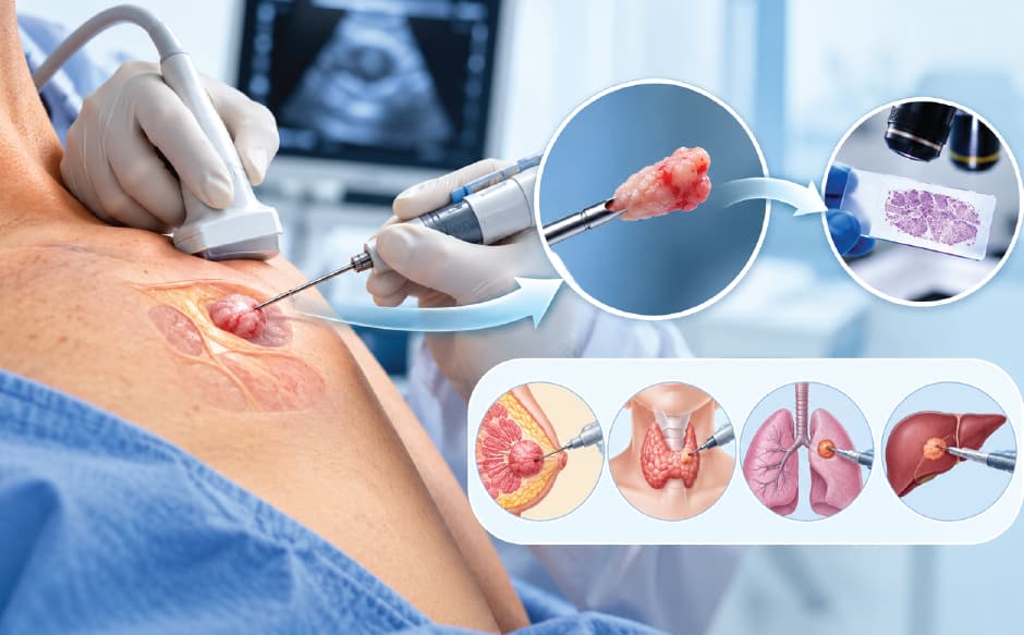
If a mole raises concern during a clinical exam, the process is usually straightforward and doesn't require assuming the worst. A dermatologist first examines the lesion with a dermatoscope, a handheld magnifying tool with polarised light that reveals pigment patterns invisible to the naked eye. If anything about the pattern looks atypical, the next step is a biopsy: either a small punch sample or a full excisional biopsy that removes the entire lesion with a narrow margin of healthy skin.
The tissue is then sent for histopathology, where a pathologist examines it under a microscope to confirm whether the cells are benign, pre-cancerous, or malignant, and if malignant, how deep the lesion has grown — a measurement called Breslow thickness that heavily influences the treatment plan. A faster, more accurate turnaround at this stage genuinely changes outcomes, since it shortens the time between "something looks off" and "here is your treatment plan."
Why NAVA Cancer Institute at BMH Is a Trusted Choice for Skin Cancer Care
So what does this actually look like if you're in Kerala and worried about a mole? At Baby Memorial Hospital, the pathway is designed to reduce the back-and-forth that can delay diagnosis and treatment. The Department of Dermatology & Cosmetology provides the initial evaluation across its Kozhikode, Kannur, Thodupuzha, Payyanur, and Vadakara locations, helping patients move from suspicion to diagnosis within the same hospital network.
A Complete Diagnostic Pathway
The initial assessment can include:
-
Clinical skin examination to assess suspicious moles or lesions.
-
Dermatoscopy for a closer, magnified evaluation of abnormal skin changes.
-
Diagnostic skin biopsy when a lesion requires tissue confirmation.
-
In-house pathology support to facilitate diagnostic reporting.
If the biopsy confirms skin cancer, the pathway continues directly into NAVA Cancer Institute, rather than requiring patients to coordinate separate oncology services elsewhere.
Multidisciplinary Cancer Care Under One Roof
NAVA Cancer Institute brings multiple specialties together, including:
-
Surgical oncology for tumour removal and surgical planning.
-
Medical oncology when systemic treatment is required.
-
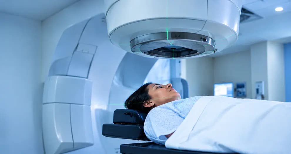
Radiation oncology when radiation forms part of the treatment plan.
-
Nuclear medicine for relevant imaging and cancer evaluation.
-
Multidisciplinary tumour board review for coordinated treatment decisions.
This team-based approach allows important decisions, such as appropriate excision margins, whether lymph node evaluation is needed, and whether systemic therapy should be considered, to be reviewed collectively before treatment is finalised.
None of this replaces your own dermatologist's advice if you already have one you trust. But if you're searching for where to get a mole checked properly in North Kerala, with dermatology, pathology, and oncology genuinely connected rather than operating as separate referrals, this is the kind of pathway worth knowing about before you need it.
Advanced Staging and Treatment Planning
For cancers such as melanoma, accurate staging can be particularly important before deciding on the extent of treatment. NAVA's diagnostic capabilities include PET-CT imaging and molecular diagnostics, which can support evaluation for disease spread and help inform treatment planning.
This integrated pathway, from dermatology assessment and biopsy through pathology, staging, tumour board review, and oncology treatment, helps minimise fragmented care.
For patients travelling from other parts of India or abroad, BMH also provides coordinated support, including treatment estimates and logistical assistance. This can make it easier to seek timely evaluation or a second opinion without allowing distance and coordination challenges to delay care.
Baby Memorial Hospital, Kozhikode was also recognised among India's Best Hospitals 2026 by Newsweek, adding another external recognition to the hospital's broader clinical profile.
If It Is Skin Cancer: Treatment Options
Treatment for confirmed skin cancer is driven almost entirely by stage at diagnosis, which is exactly why early detection matters as much as it does.
| Stage / Situation | Typical Treatment Approach |
|---|---|
| Very early, thin lesion | Wide local excision under local anaesthesia, often curative on its own |
| Deeper or higher-risk lesion | Wider surgical margins plus sentinel lymph node biopsy to check for spread |
| Lymph node involvement | Surgical clearance combined with adjuvant immunotherapy or targeted therapy |
| Advanced or recurrent disease | Systemic immunotherapy, targeted drugs, and/or radiotherapy, guided by a tumour board |
Most skin cancers, including a large share of melanomas caught through routine self-exams, are treated successfully with surgery alone. The more advanced options exist for the smaller number of cases caught later or that have already spread, which is precisely the scenario the ABCDE rule and regular self-checks are designed to help you avoid.
A Simple Monthly Self-Exam You Can Start Today
-
Use a well-lit room and a full-length mirror, with a hand mirror for hard-to-see areas like your back, scalp, and soles.
-
Work in a consistent order, face, scalp, neck, arms, torso, legs, feet, and between fingers and toes, so you don't skip areas.
-
Take a photo of any mole you're unsure about, so you have an objective reference point a month later.
-
Note the date you check, so "evolution" changes are measured in weeks and months, not guesswork.
-
If anything matches two or more ABCDE signs, don't wait for your next scheduled check, book a dermatology appointment.
Conclusion
Most moles you'll ever notice on your skin are, and will remain, completely harmless. But the small minority that aren't follow a fairly predictable pattern, asymmetry, irregular borders, uneven colour, growing diameter, and ongoing change, that the ABCDE rule was built to catch early, before a lesion has the chance to spread.
The single most useful habit you can build is a simple one: check your skin monthly, know your own moles well enough to spot the "ugly duckling," and treat any change as a reason to get a professional opinion rather than a reason to wait.
If that opinion is ever needed in Kerala, BMH's dermatology team and NAVA Cancer Institute are set up to take you from a first look to a confirmed diagnosis and, if necessary, a full treatment plan, without losing time between steps.
Chat with our medical assistant on the Baby Memorial Hospital for guidance, or book a dermatology consultation at your nearest BMH location.
Medical Disclaimer: This article is intended for general informational purposes only and does not constitute medical advice, diagnosis, or treatment. Always consult a qualified dermatologist or oncologist for evaluation of any mole, skin lesion, or symptom you are concerned about. Baby Memorial Hospital and NAVA Cancer Institute do not assume responsibility for decisions made based solely on this content.