
Most women feel alarmed when they discover a breast lump, fearing an immediate cancer diagnosis. The truth is that the vast majority of breast lumps are benign, non-cancerous growths that require monitoring but not aggressive treatment. Understanding the key differences between benign lumps and breast cancer helps you stay informed and take the right steps without unnecessary panic.
What Exactly Is a Breast Lump?
Breast tissue is not uniform. It's a mix of glandular tissue, fat, and connective tissue that changes texture with your menstrual cycle, age, pregnancy, and hormone levels. Some people naturally have "lumpier" or denser breasts than others, and knowing your own baseline is more useful than comparing yourself to a textbook description.
Most new lumps trace back to benign (non-cancerous) causes, including:
- Fibroadenomas, the most common benign lump, typically smooth, rubbery, and mobile
- Breast cysts, fluid-filled sacs, often tender and more noticeable before a period
- Fibrocystic changes, widespread lumpiness tied to hormonal cycles, affecting as many as half of all women at some point
- Mastitis, an infection, usually during breastfeeding, causing a warm, tender, swollen area
- Lipomas, soft, painless fatty growths
None of these are cancer. A few, like certain fibrocystic patterns, are associated with a slightly higher future risk, which is a separate conversation from "is this lump cancer right now."
Breast Lump vs Breast Cancer: Side-by-Side Comparison
Doctors look at several physical characteristics together, not any single one in isolation. Here's how they generally differ, with the important caveat that overlap exists, and touch alone is never a diagnosis.
| Characteristic | Typically Benign | Watch Closely / See a Doctor |
|---|---|---|
| Texture Soft, smooth, or rubbery | Hard, like a marble or pebble | |
| Borders | Well-defined, rounded | Irregular, poorly defined |
| Mobility | Moves freely under the skin | Fixed in place, doesn't shift when pressed |
| Pain | Often tender, changes with menstrual cycle | Usually painless (though not always) |
| Growth pattern | Stable or fluctuates with cycle | Grows steadily over weeks |
| Skin/nipple changes | None | Dimpling, puckering, nipple retraction, or discharge |
| Associated swelling | None | Underarm or collarbone swelling |
Even breast surgeons are candid that texture alone can mislead, a cancerous lump can occasionally feel soft, and a benign cyst can feel unusually firm if it sits deep in the tissue. That's precisely why "does it feel hard" is a starting question for a clinician, not an answer for a patient sitting at home.
Warning Signs That Need Same-Week Medical Attention
Certain signs shift the picture from "monitor it" to "get seen this week," regardless of how the lump itself feels:
- A lump that feels hard and doesn't move when you press around it
- Skin dimpling or a texture resembling an orange peel
- A nipple that suddenly turns inward or changes direction
- Clear or bloody nipple discharge not caused by squeezing
- New swelling under the arm or near the collarbone
- Any change that persists beyond one full menstrual cycle
How doctors tell the difference between benign lumps and cancer
Clinicians don't rely on appearance or feel alone. The standard approach, often called a triple assessment, combines three independent checks, because each one catches something the others might miss.
1. Clinical Breast Examination
A physician manually examines both breasts and the lymph node regions (armpit, collarbone), checking texture, mobility, size, and symmetry, alongside a detailed history of when the lump appeared and any family history of breast or ovarian cancer.
2. Imaging
Mammography, an X-ray of breast tissue, generally the first imaging step, especially for women over 40 Ultrasound, often used alongside or instead of mammography for younger women or denser breast tissue, and to determine whether a lump is solid or fluid-filled MRI, reserved for specific higher-risk cases or when mammogram and ultrasound findings are unclear.
3. Biopsy
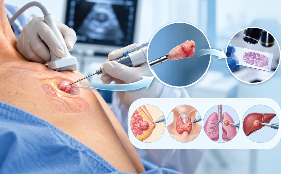
If imaging flags anything suspicious, a small tissue sample is taken, usually via fine-needle aspiration cytology (FNAC) or a core needle biopsy, and examined under a microscope. This is the only step that gives a definitive answer. Everything before it narrows the possibilities; only a biopsy confirms them.


How NAVA Cancer Institute at Baby Memorial Hospital Diagnoses and Treats Breast Lumps
At NAVA Cancer Institute, part of Baby Memorial Hospital (BMH) in Kozhikode, the triple assessment isn't spread across separate appointments and separate facilities, clinical evaluation, imaging, and biopsy are coordinated within the same centre, which shortens the anxious waiting period between "something feels different" and "here's what it actually is."
Imaging is done on BMH's Siemens MAMMOMAT Revelation, a digital mammography system built to detect breast tissue changes at an early stage, often before they're physically noticeable, a meaningful advantage for women whose lump turns out to be nothing, since it rules out cancer with more confidence than a physical exam alone, and equally meaningful for the smaller group whose lump is malignant, since earlier detection directly improves treatment options.
Breast cancer cases at NAVA are managed by a multidisciplinary team, with the Medical Oncology team comprising Dr. Muhammad Rifayi V, Dr. Akhil Santhosh, and Dr. Nithin Raj; surgical support from Dr. John J. Alapatt and Dr. Sanoor Neha Kamath, and the Radiation Oncology team comprising Prof. Dr. P. R. Sasindran, Dr. Dhanya K. S, Dr. Midhun Murali and Dr. Vineeth Mathew John. Depending on the stage and type of cancer, treatment can involve breast-conserving surgery (lumpectomy), mastectomy, chemotherapy, radiation therapy, targeted therapy, or hormone therapy, sequenced according to the specific case rather than a fixed protocol.
What to Do If You Find a Lump: Step by Step
- Note the details, size, location, when you first noticed it, whether it changes with your cycle
- Resist the urge to self-diagnose from symptom checklists, they narrow possibilities, they don't confirm anything
- Book a clinical breast exam within days, not weeks, most causes are benign, but timing matters for the minority that aren't
- Bring your family history, first-degree relatives with breast or ovarian cancer changes the risk calculus and the recommended screening approach
- Follow through on any imaging or biopsy recommended, an incomplete workup is often worse than the anxiety of getting the full picture.
Also Read: Hormone Therapy For Breast Cancer: Complete Guide
Conclusion
Finding a breast lump is unsettling, and the instinct to fear the worst is understandable, but the evidence doesn't support that fear as the likely outcome. Most lumps are benign. What separates a good outcome from a delayed diagnosis isn't how the lump feels under your fingers; it's whether you get it properly assessed through clinical examination, imaging, and biopsy when needed, rather than relying on guesswork either way. Texture, mobility, and pain can offer early clues, but only a triple assessment gives a real answer, and the earlier that assessment happens, the more treatment options remain on the table if it does turn out to be cancer. Whether the lump is nothing to worry about or something that needs treatment, timely evaluation is the one variable you fully control.
You don't need to sit with uncertainty. Chat with our assistant to understand next steps based on what you're experiencing, or book a consultation with NAVA Cancer Institute at Baby Memorial Hospital for a same-facility clinical exam, imaging, and biopsy pathway if needed.