
Melanoma is one of the most dangerous forms of skin cancer, but it is highly treatable when caught early. The ABCDE rule is a simple framework that helps you and your doctor identify suspicious moles before they become life-threatening. By learning to recognize these five warning signs, you can take charge of your skin health and seek timely care when needed.
What Is Melanoma, and Why Does Early Detection Matter?
Melanoma begins in melanocytes, the cells that produce the pigment (melanin) responsible for skin, hair, and eye colour. Unlike the two more common forms of skin cancer (basal cell and squamous cell carcinoma), melanoma is far more likely to spread to lymph nodes and internal organs if it isn't caught early, which is why it accounts for the majority of skin cancer deaths despite being a smaller share of overall skin cancer cases.
A normal mole (nevus) tends to be a stable, unremarkable part of your skin, round, evenly coloured, and roughly the same for years. Melanoma tends to break at least one of those patterns. It rarely announces itself with pain in the early stages, which is precisely why a visual checklist matters so much: it gives you something concrete to look for before symptoms like bleeding or ulceration appear, by which point the cancer has often had time to grow deeper into the skin.
Dermatologists Darrell Rigel, Robert Friedman, and Alfred Kopf first proposed the ABCD rule in the 1980s, and it was later expanded to ABCDE to capture change over time, arguably the single most useful clue a self-exam can offer.
The ABCDE Rule for Melanoma Detection, Explained
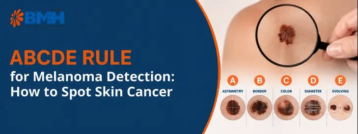
The ABCDE rule gives you a simple way to assess moles and other pigmented spots for changes that may need a closer look. Each letter represents a warning sign to check during a skin self-exam.
1. A — Asymmetry
Draw an imaginary line through the middle of a mole. In a benign mole, both halves generally match. In melanoma, one half often looks noticeably different from the other in shape or contour, one side may bulge, taper, or extend further than the other.
2. B — Border
Healthy moles usually have smooth, even edges. Melanoma borders are frequently irregular, notched, scalloped, or poorly defined, sometimes fading unevenly into the surrounding skin rather than stopping cleanly.
3. C — Colour
A benign mole is typically one consistent shade of brown. Melanoma often contains a mix of colours within the same lesion, different shades of tan, brown, or black, and sometimes patches of red, white, or blue-grey.
4. D — Diameter
Melanomas are often larger than 6 millimetres across, roughly the size of a pencil eraser, when diagnosed, though they can be caught smaller. Diameter alone is the least reliable letter in the rule; it's best read alongside the others.
5. E — Evolution
Any change, in size, shape, colour, elevation, or a new symptom like itching, tenderness, or bleeding, is often considered the most important warning sign of all. A mole that looked stable for years and then starts changing over weeks or months deserves a professional look, even if it doesn't tick every other box.


Beyond ABCDE: Signs the Rule Can Miss
The ABCDE rule is a screening tool, not a diagnosis, and it isn't foolproof. A meaningful share of melanomas, particularly nodular melanoma (which grows downward rather than outward) and amelanotic melanoma (which has little or no pigment), don't fit the classic asymmetric, multicoloured pattern at all. A nodular melanoma can be a small, firm, symmetrical, single-coloured bump that grows quickly, and still be dangerous.
This is why dermatologists also teach the "ugly duckling" sign: if one mole on your body looks distinctly different from all your other moles, not necessarily large, dark, or irregular, just different, it's worth having checked, even if it passes every ABCDE letter individually.
Alongside appearance, pay attention to a spot that starts to itch persistently, bleed without an obvious injury, or develop a crust that doesn't heal. None of these are signs to self-diagnose, but they are reasons to book an appointment rather than wait for the next self-exam.
How to Do a Skin Self-Exam at Home (Step-by-Step)
A skin self-exam takes only a few minutes, but doing it systematically helps you notice new or changing spots that might otherwise be easy to miss. Follow these steps from head to toe:
-
Pick good lighting and two mirrors: A full-length mirror and a hand-held mirror for hard-to-see spots.
-
Work systematically, front to back. Face, ears, neck, and scalp (a comb or hairdryer helps part hair). Chest, torso, and, for those with breasts, the underside.
-
Raise your arms and check underarms, then your arms front and back, down to your palms and between your fingers.
-
Sit down to check your legs, feet, soles, and between your toes. These areas are often missed but are common sites for melanoma, especially in people with darker skin tones.
-
Use the hand mirror for your back, buttocks, and genitals, or ask a partner to help.
-
Photograph anything you're unsure about, dated, so you have a record to compare against next time.
-
Repeat monthly, and schedule a professional skin check with a dermatologist at least once a year, more often if you have many moles, fair skin, or a family history of melanoma.
What Happens After You Spot a Suspicious Mole: Diagnosis & Staging
If a mole raises concern, a dermatologist will typically start with dermoscopy, examining the lesion under magnification with polarised light to look at pigment patterns invisible to the naked eye.
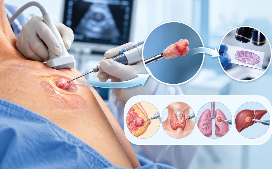
If it still looks suspicious, the next step is a biopsy, usually an excisional biopsy that removes the entire lesion with a small margin of healthy skin for lab analysis.
If melanoma is confirmed, staging determines how far it has spread. This typically involves imaging such as a PET-CT scan and, depending on the tumour's thickness, a sentinel lymph node biopsy to check whether nearby lymph nodes are involved.
Staging is what shapes every treatment decision that follows, which is why getting from "suspicious mole" to "confirmed stage" quickly, under one coordinated team, matters as much as the diagnosis itself.
Why Patients Across India Choose NAVA Cancer Institute at Baby Memorial Hospital for Cancer Care
If a mole is worrying you or someone in your family, a melanoma diagnosis or even the possibility of one, involves more than just a medical decision. Patients also need to consider where to get a biopsy, who will review the pathology, and whether diagnosis and treatment can be coordinated without moving between multiple hospitals.
NAVA Cancer Institute at Baby Memorial Hospital (BMH), Kozhikode, brings different areas of cancer care together within one integrated setting. This can help patients move more smoothly from initial evaluation and biopsy to staging, treatment planning, and follow-up.
What Makes NAVA Cancer Institute a Practical Choice?
-
Multidisciplinary cancer care: Medical oncology, surgical oncology, radiation oncology, and nuclear medicine work together through a multidisciplinary tumour board to review cases and develop an appropriate treatment plan.
-
Integrated diagnostic services: Imaging, pathology, and oncology services are available within the hospital, helping reduce the need to coordinate investigations across separate centres.
-
Advanced cancer imaging: Facilities such as Discovery IQ PET-CT can support staging and treatment planning when clinically indicated.
-
Surgical expertise and technology: For selected patients who require surgery, minimally invasive and robotic approaches, including Da Vinci robotic surgery, may be considered when appropriate.
-
In-house pathology: Biopsy specimens can be evaluated within the hospital, helping streamline the diagnostic process.
-
Support for patients travelling from outside Kerala: Dedicated patient coordination services can help patients and families manage appointments, investigations, and treatment planning.
-
International patient support: Patients travelling from abroad can access assistance with coordination and communication, including multilingual support.
For patients travelling from other parts of India or internationally, having multiple cancer services coordinated within one hospital can make the treatment journey more manageable. Instead of arranging separate consultations and investigations at different centres, patients can have their case evaluated through an integrated cancer-care pathway.
For melanoma specifically, however, the most important factor is timely and accurate diagnosis followed by treatment appropriate to the stage and characteristics of the cancer. The choice of hospital should therefore be based on the availability of the right specialists, pathology expertise, imaging, surgical options, and multidisciplinary decision-making.
NAVA Cancer Institute at BMH provides these services within a coordinated cancer-care setting, allowing patients to progress from suspicious mole evaluation and biopsy to staging and treatment planning without unnecessarily fragmenting their care.
The information above is intended for general awareness. A dermatologist or qualified cancer specialist should evaluate any changing, suspicious, or unusual mole.
Melanoma Treatment Options at a Glance
Treatment depends almost entirely on stage:
-
Surgical excision is the primary treatment for early-stage melanoma, removing the tumour with a margin of healthy skin.
-
Sentinel lymph node biopsy may be done alongside surgery for thicker tumours, to check for early spread.
-
Immunotherapy and targeted therapy have substantially changed outcomes for melanoma that has spread regionally or to distant sites, using drugs that either help the immune system recognise cancer cells or block the specific mutations driving a tumour's growth.
-
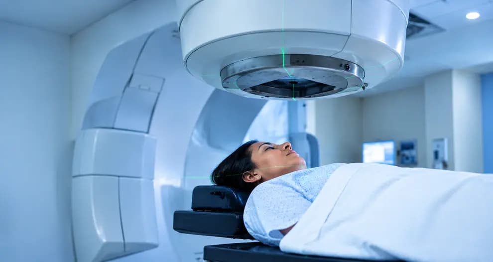
Radiation therapy is used selectively, often for tumours in difficult-to-operate locations or to manage spread to lymph nodes or other sites.
A tumour board reviews all of this together, pathology, imaging, and the patient's overall health, before recommending a plan, which is standard practice at comprehensive cancer centres rather than something unique to any one hospital.
Conclusion
The ABCDE rule won't diagnose melanoma, only a dermatologist and a biopsy can do that, but it gives you something far more valuable in the meantime: a reason to look, and a reason to act early rather than wait and watch.
Asymmetry, an irregular border, uneven colour, a growing diameter, and above all, evolution over time, are the five signals worth building into a monthly habit. Combine that with an annual professional skin check, and you're catching melanoma at the stage where it's most treatable, not after it's had time to spread.
If a mole on your skin doesn't look quite right, that's reason enough to get it looked at, regardless of how many ABCDE boxes it ticks.
If a spot on your skin has you wondering, don't wait for it to change further. Contact NAVA Cancer Institute at Baby Memorial Hospital, Kozhikode to arrange a dermatology consultation, or chat with our assistant on Whatsapp to ask a quick question and get pointed to the right specialist.
Medical Disclaimer: This article is for general informational purposes only and does not constitute medical advice, diagnosis, or treatment. Always consult a qualified dermatologist or oncologist for evaluation of any skin changes or symptoms. Baby Memorial Hospital and NAVA Cancer Institute do not guarantee specific outcomes; individual results vary by diagnosis, stage, and patient health.